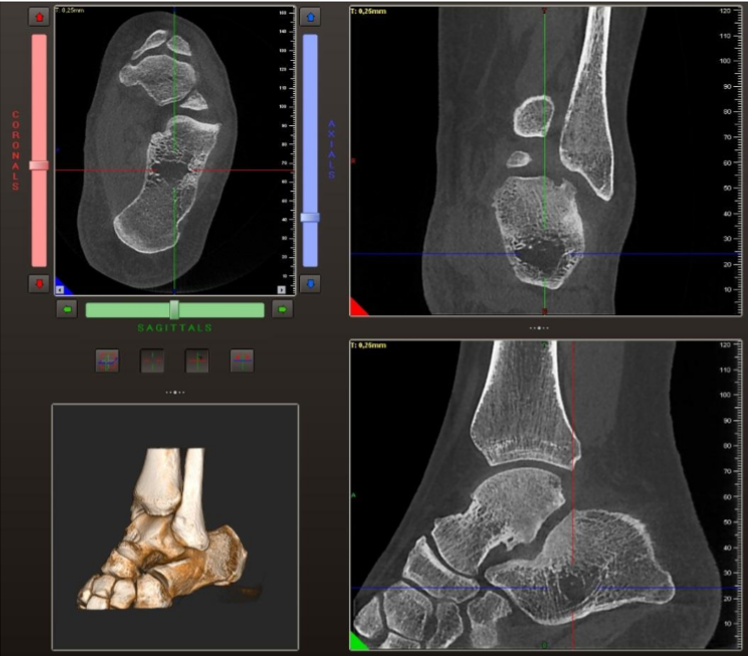
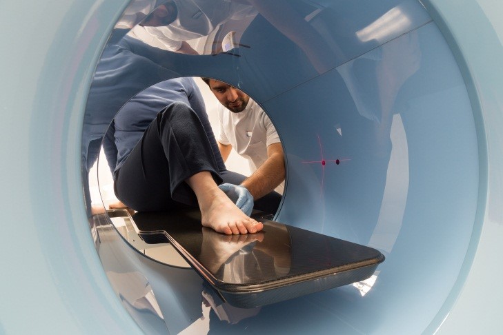
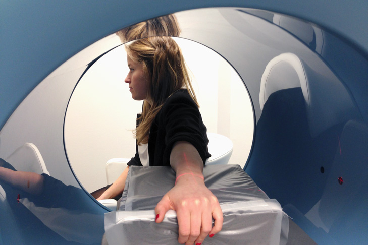
Cone-beam computed tomography (CBCT) is continually being proven useful for orthopaedic cases and especially when using the Newtom 5G scanner. This CBCT scanner allows a higher degree of free positioning of the patient and therefore no longer limits the use of CBCT to the dentomaxillofacial region.
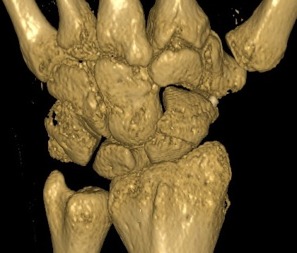
In one instance, CBCT was shown to be useful in the diagnosis of a wrist injury. A CBCT scan was able to show a small 1 mm residual intra-articular step-off that could not be seen on a conventional radiograph. In the traditional radiograph, the wrist actually appeared normal, despite the patient’s decreased range of motion and persisting pain.
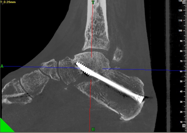
In another case, a patient had continuing pain after having a scaphoid fracture treated with a Herbert screw. A CBCT scan was able to show that the screw was not well incorporated into the distal pole of the scaphoid. Traditional radiographs were only able to show that it was well incorporated in the proximal pole of the bone. The metal scattering artefact from the screw may have hindered mal-union diagnostic. CBCT helped patients receive better care in both of these cases.
Limiting the radiation dose to the patients must also be everyone’s objective. The radiation delivered by our CBCT scanner is 65-92% less than a conventional CT (Smith et al). The radiation received by the patient from an ankle CBCT using our standard protocol is 10 times less than that of a conventional CT scanner and equivalent to that of 2 x-rays (Koivisto et al) but, with obviously, the whole 3D information.


