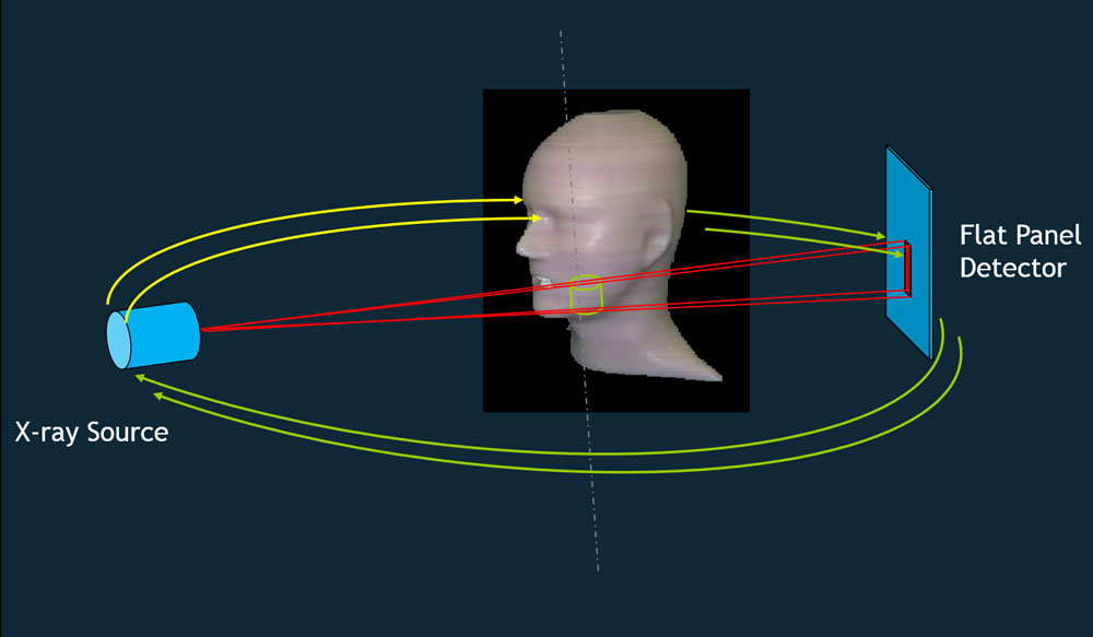
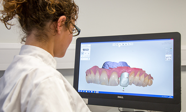
Cone Beam CT (CBCT) is an X-ray based imaging technique that, like a conventional medical CT scan, provides fast and accurate visualisation of bony anatomical structures in three dimensions.
The critical difference with CBCT scanning is that it produces higher resolution images, with less artefact and scatter, making it the scanning technique of choice for the imaging of hard tissues. Additionally, the scan field of view can also be reduced to image smaller volumes.
We select our scanners based on the radiation optimisation facilities that they give us and the image quality they produce for a given radiation dose. Our high-end CBCT scanners also offer great flexibility on many scanning parameters. For instance, we can reduce volume sizes and acquire the data of the area of interest through a single or partial rotation of the conical x-ray beam and reciprocal image receptor. All of this contributes to a lower radiation dose associated with each scan.
The high detail of our smallest scan volumes makes our CBCT scanners a powerful diagnostic tool in finding small fractures very quickly. The immediate production of skeletal 3D anatomy also hugely expands diagnostic and treatment possibilities for your patients.
Our scanning protocols are carefully developed. We work in close collaboration with academia, NHS teaching hospitals, regulators and equipment manufacturers to optimise our image quality for each application, which ranges from dentistry to craniomaxillofacial and ENT (ear, nose and throat) applications as well as orthopaedics.


