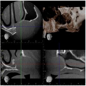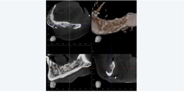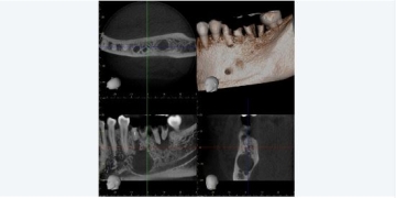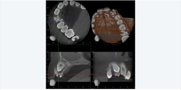
CBCT showing the ULQ with edentulous left maxilla. There is a well defined expansile left maxillary lucent bone lesion.
The appearances and expansile nature in absence of residual dentition mean a radicular cyst is unlikely. The degree of expansion can be seen with maxillary KCOT, but no internal scalloping is perhaps atypical – but KCOT is not excluded.
Alternative diagnoses include Giant Cell Tumour (although age group is atypical), Aneurysmal Bone Cyst, Plasmacytoma or less likely a metastasis (in the absence of any prior medical history).
Surgical biopsy was recommended – and any further imaging (eg staging scans / bone scan) would depend upon the histological result of biopsy.