Our high-end Cone Beam CT (CBCT) scanners are proving their capability for temporal bone exploration.
Referring criteria: anatomical variants, dysplasia, chronic otitis, deformity, trauma, pre and post-cochlear implantation.
Imaging: JMorita Accuitomo F170 and NewTom 5G
Radiation dose: Typically between 150mSv – 250mSv (approx. 20-40% of conventional CT)
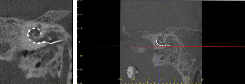
Findings: Case Studies 1-6 below highlight our CBCT image quality and its appropriateness over CT. CBCT’s low sensitivity to metallic artefacts makes it the technique of choice in the follow up of cochlear implants. The significantly lower radiation doses delivered using CBCT in relation to CT complement guidance for best clinical practice.
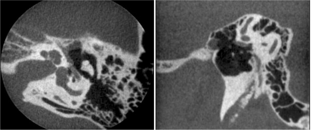
The Newtom 5G 15x5cm FOV is proving to be diagnostically advanced when imaging temporal bones with high depiction and differentiation of the inner and middle ear complexes.
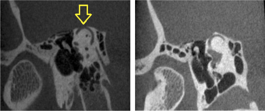
There is dehiscence of the right superior semi-circular canal over a 3.5mm distance (see yellow arrow). The left canal lies very close to the cortical margin but it remains intact.
CBCT is preferable for the follow up of cochlear implant investigation due to the lower rate of metallic artefact. See the example below for high resolution views.
Post tympanoplasty surgery, the images below confirm the correct placement of the prosthesis (green arrow). The yellow arrow indicates the thinned facial nerve canal. The red arrow highlights the residual head of the hammer.
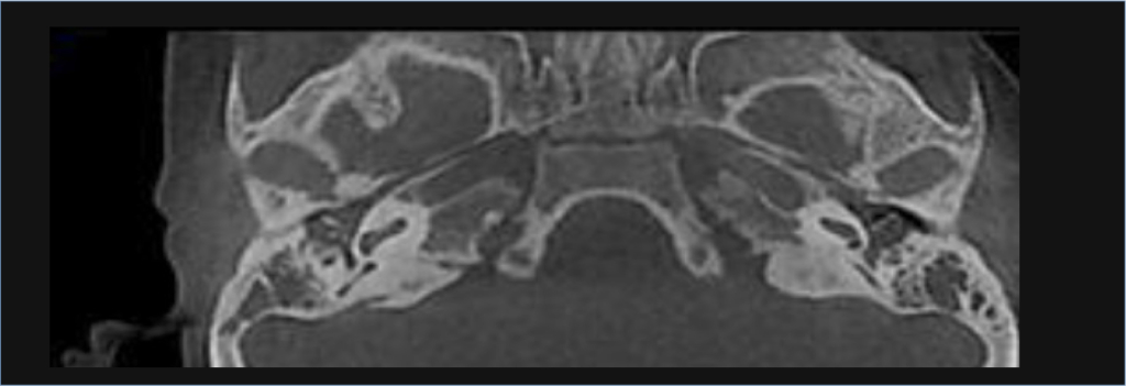
The axial image below shows clearly the eustachian tubes with bilateral otitis media.
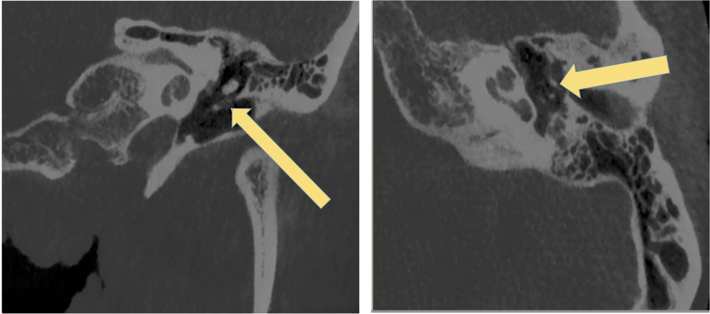
Non-specific abnormal soft tissue lining the anterior wall of the right epitympanic.
Bilateral minor thickening and faint calcification of the posterior aspect of both tympanic membranes indicated with yellow arrows below.

